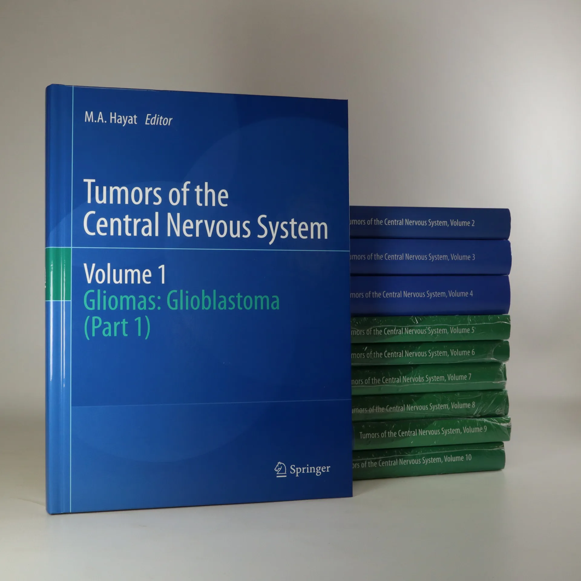
This book presents recent developments in diagnostic and therapeutic aspects of Gliomas (Glioblastoma) in the brain. It examines the importance of personalized medicine and clinical validation for targeted therapy.
Mohammad Hayat Book order (chronological)
January 1, 1940
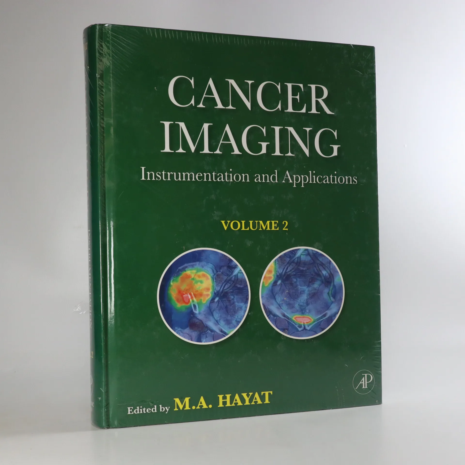
Cancer Imaging
- 656 pages
- 23 hours of reading
These two volumes present readers with the most up-to-date imaging instrumentation, general and diagnostic applications for various cancers. Both volumes discuss the various imaging techniques used to locate and diagnose tumors, including ultrasound, X-ray, color Doppler sonography, PET, CT, PET/CT, MRI, SPECT, diffusion tensor imaging, dynamic infrared imaging, and magnetic resonance spectroscopy. They also detail strategies for imaging cancer, emphasizing the importance of the use of this technology for clinical diagnosis. Imaging techniques that predict the malignant potential of cancers, response to chemotherapy and other treatments, recurrence, and prognosis are also detailed.
This second of two volumes on Cancer Imaging covers the three major topics of imaging instrumentation, general imaging applications, and imaging of a number of human cancer types. Where the first volume emphasized lung and breast carcinomas, Volume 2 focuses on prostate, colorectal, ovarian, gastrointestinal, and bone cancers. Although cancer therapy is not the main subject of this series, the crucial role of imaging in selecting the type of therapy and its post-treatment assessment are discussed. The major emphasis in this volume is on cancer imaging; however, differentiation between benign tumors and malignant tumors is also discussed. This volume is sold individually, and Cancer Imaging, Volume 1 [ISBN: 978-0-12-370468-9] sells separately for $189 and also as part of a two volume set [ISBN: 978-0-12-374212-4] for $299. • Concentrates on the application of imaging technology to the diagnosis and prognosis of prostate, colorectal, ovarian, gastrointestinal, and bone cancers • Addresses relationship between radiation dose and image quality • Discusses the role of molecular imaging in identifying changes for the emergence and progression of cancer at the cellular and/or molecular levels
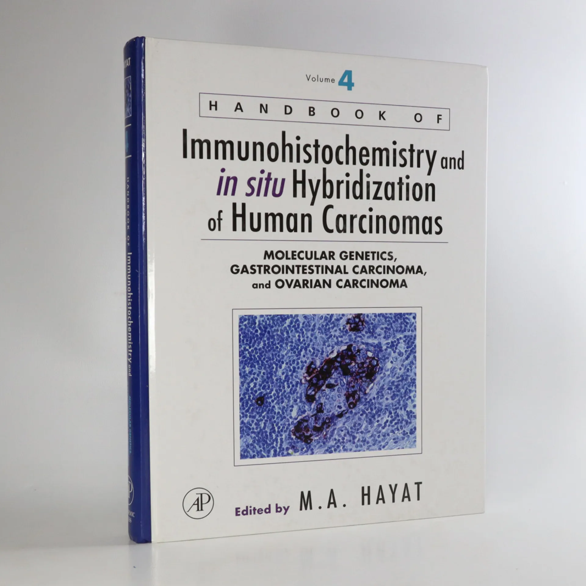
Handbook of Immunohistochemistry and in Situ Hybridization of Human Carcinomas 4
- 608 pages
- 22 hours of reading
Translating molecular genetics into cancer diagnosis, this book discusses various aspects of immunohistochemistry and in situ hybridization technologies and the important role they play in reaching a cancer diagnosis. It also provides step-by-step instructions on the methods of additional technologies such as DNA microarrays, and microdissection.
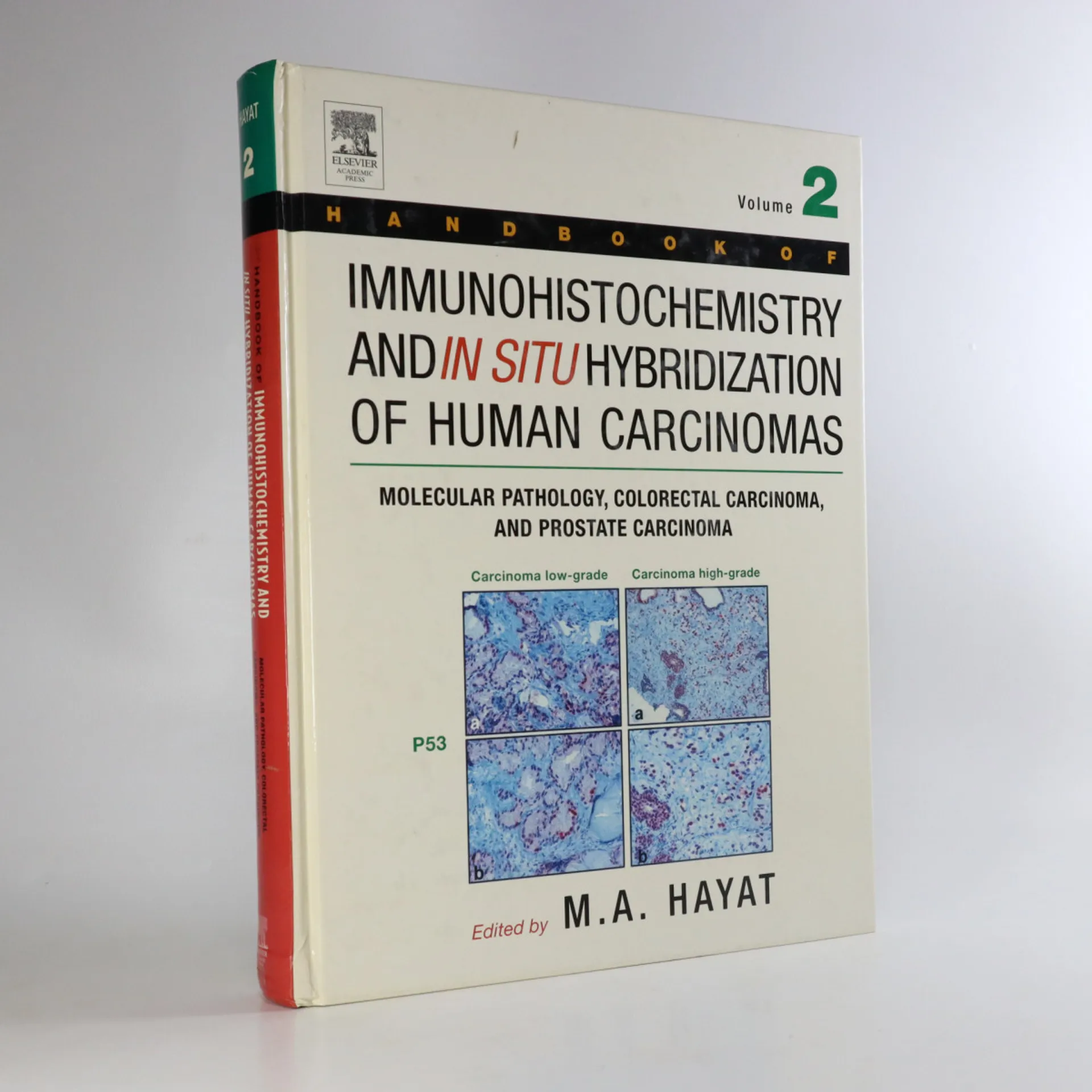
Handbook of Immunohistochemistry and in Situ Hybridization of Human Carcinomas 2
- 492 pages
- 18 hours of reading
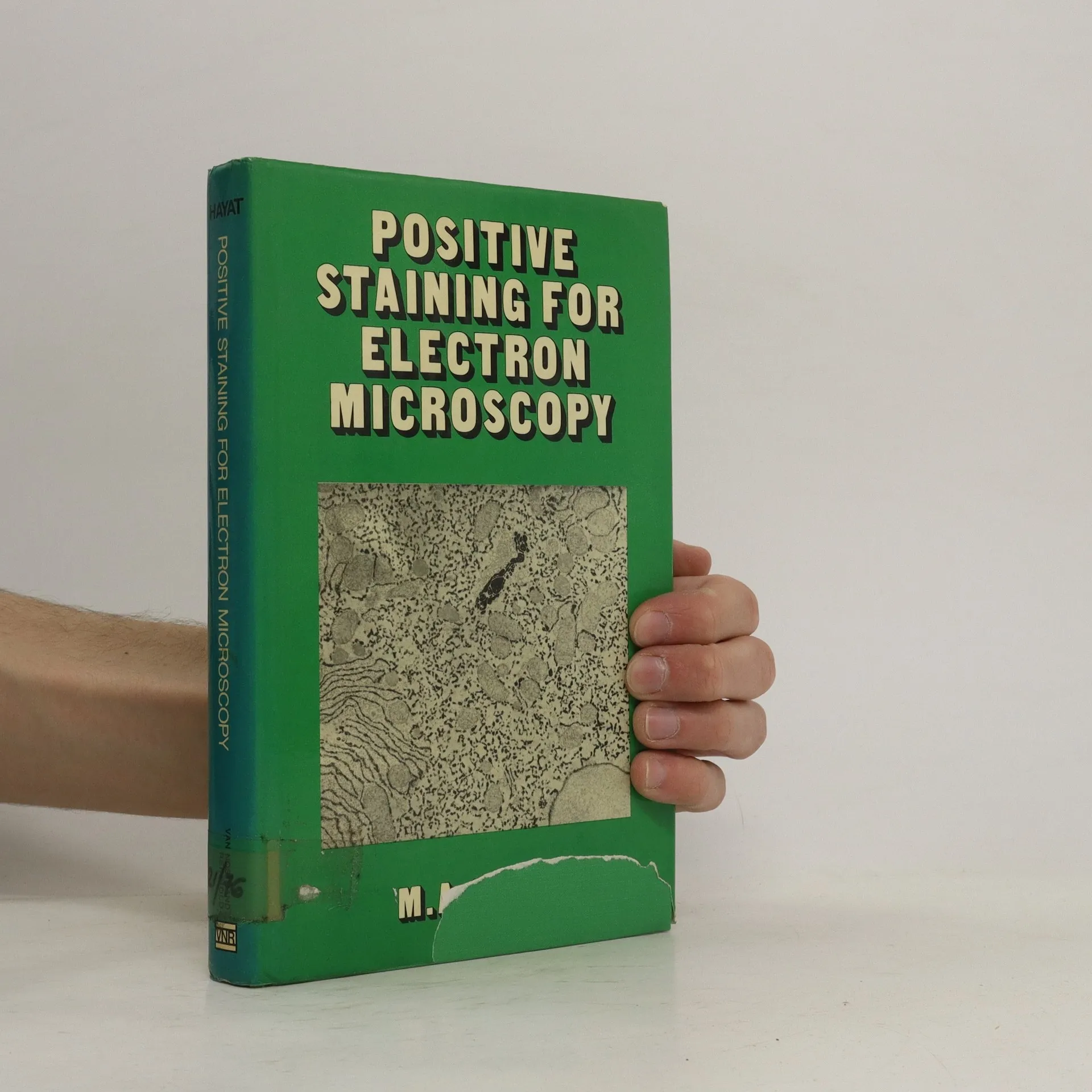
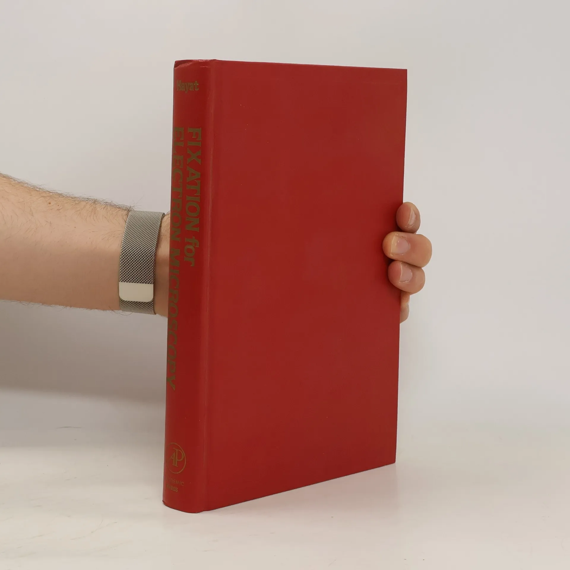
Fixation for Electron Microscopy
- 501 pages
- 18 hours of reading
Fixation for Electron Microscopy presents how to better understand the effects of fixatives on the molecular structure of the cell. This book attempts to consider each aspect of fixation, including chemical interactions between fixatives and individual cellular substances. The chemistry of fixative interactions that are discussed in the book is based primarily on the reactions of a fixative with isolated proteins, lipids, nucleic acids, and carbohydrates. The book shows that the correct interpretation of information retrieved from electron micrographs depends on the knowledge of the basic principles underlying the fixation procedure. Also, the book presents the fixation of both eukaryotic and prokaryotic specimens. The special fixation conditions for plant specimens are discussed in detail and have been allotted a whole chapter. Also emphasized in this book is the connection between morphology and biochemical aspects of preparatory treatments and the chemical basis of the formation of artifacts. This topic is useful in understanding the modifications of cell structures introduced during their processing. A guide for recognizing and minimizing major artifacts and fixation faults that are usually encountered is also presented in the book. This valuable resource will prove useful to both students and professionals in the field of biology and clinical medicine. Specimen preservation researchers can also benefit from this book.
361p cloth with green dustjacket, spine sunned, inscription to endpaper, pages unmarked, binding firm, very good, this copy published in the year 1975
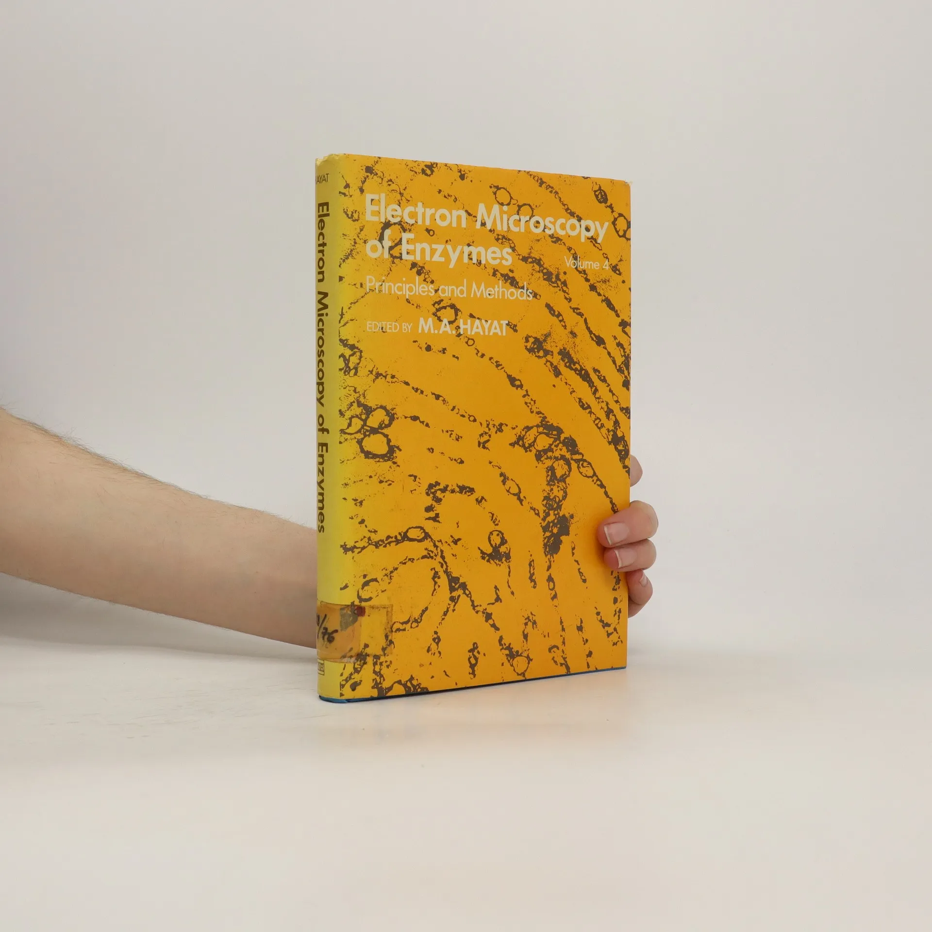
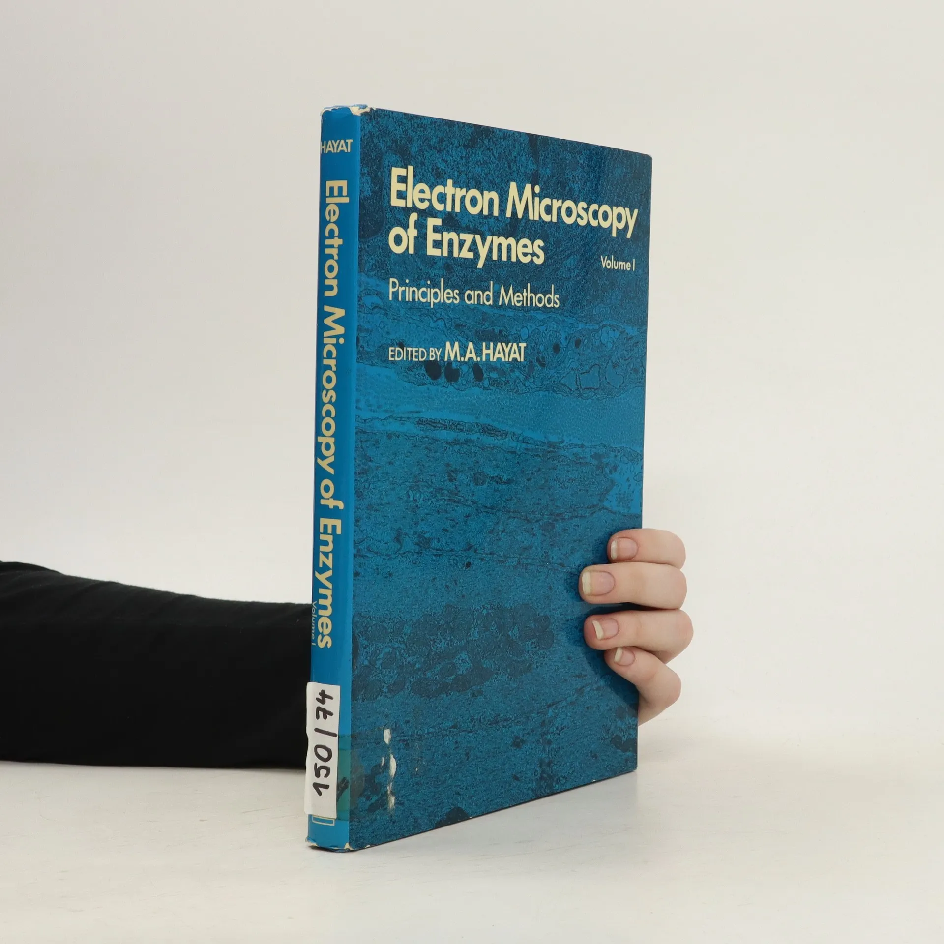
Electron Microscopy of Enzymes
- 158 pages
- 6 hours of reading
homoproteins, diaminobenzideine-peroxidase; engogenous hydrogen peroxide; adsorption of DAB, oxidized DAB, catalase, exogenous hemoproteins, cytocrhome c, cytochemical reaction, mercaptide formation, oxidation of free CoA, carnitine acetyltransferase, intestinal lipid absorption, tyrosinases, ortho-dephenoloxidases, para-diphenoloxidases, DOPA, autooxidation, melanin granules, arylsulfatases, 4-methylcatechol, parenchyumal, reticuloenthothelial cells, Islets of Langerhans, capillary endothelial cells, rod outer segments, photoreceptor membranes, lipase, nonspecific lead depositions, lead nitrate solution; nonspecific esterases; etc., etc. Black & White photographs. 158 pages.
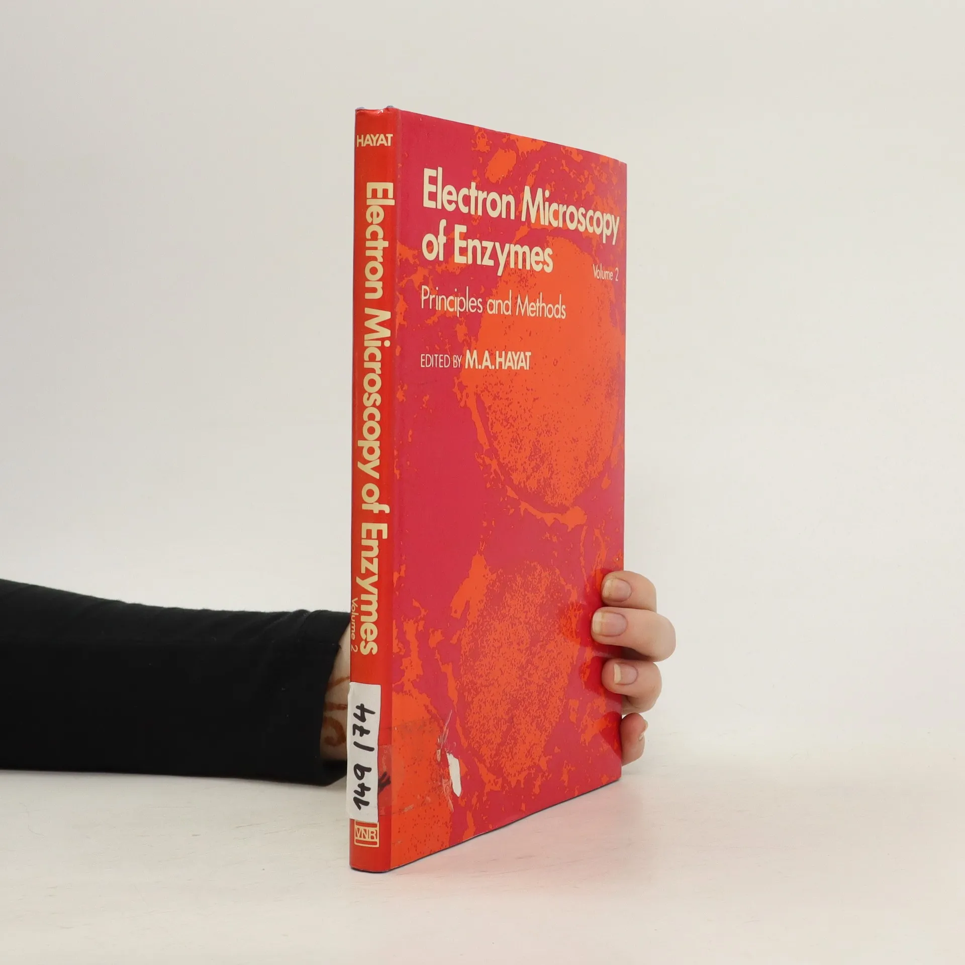
Electron Microscopy of Enzymes vo. 4
- 200 pages
- 7 hours of reading