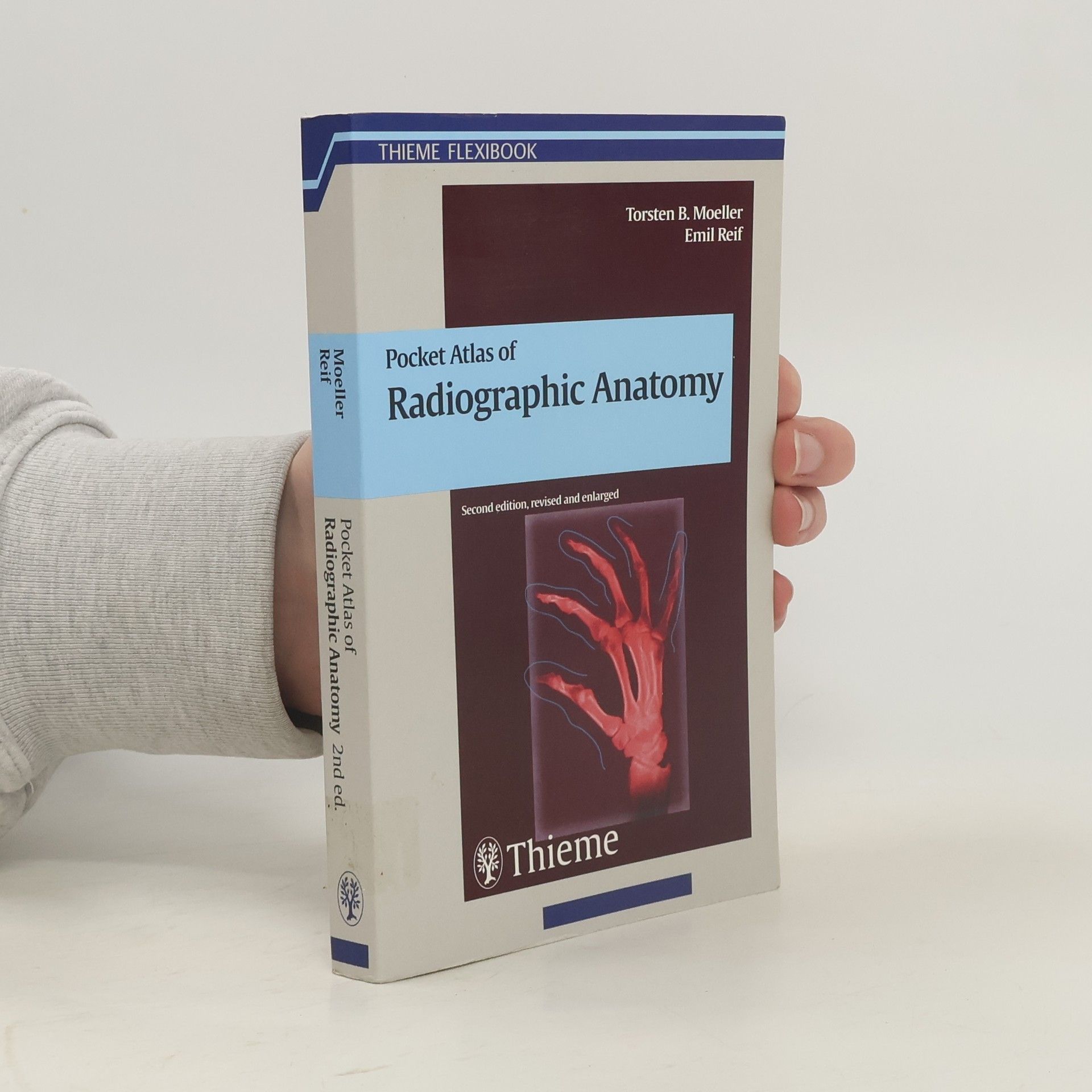
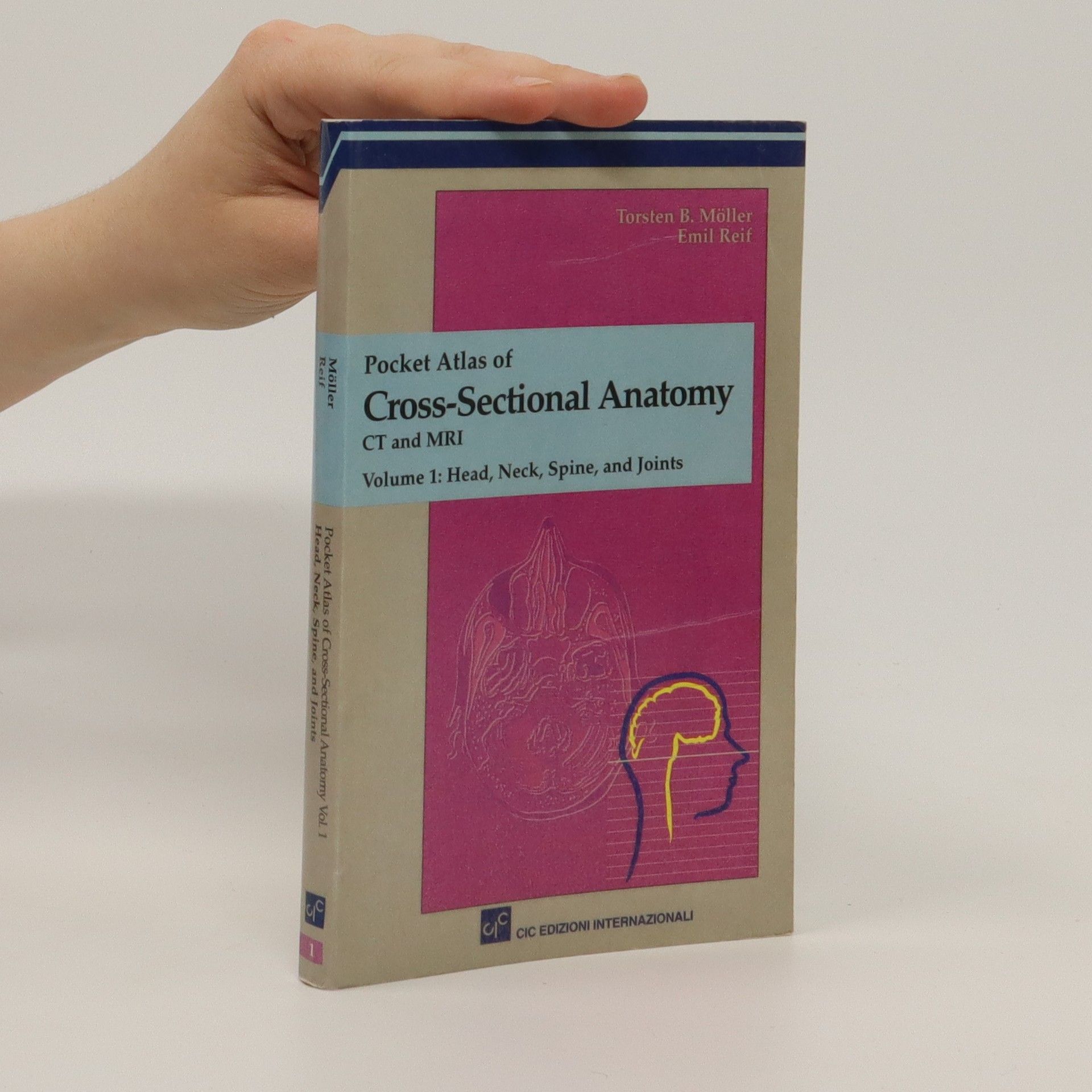
This comprehensive, easy-to-consult pocket atlas is renowned for its superb illustrations and ability to depict sectional anatomy in every plane. Together with its two companion volumes, it provides a highly specialized navigational tool for all clinicians who need to master radiologic anatomy and accurately interpret CT and MR images. Special features of Pocket Atlas of Sectional Anatomy: Didactic organization in two-page units, with high-quality radiographs on one side and brilliant, full-color diagrams on the other Hundreds of high-resolution CT and MR images made with the latest generation of scanners (e. g., 3T MRI, 64-slice CT) Consistent color coding, making it easy to identify similar structures across several slices Concise, easy-to-read labeling of all figuresUpdates for the 4th edition of Volume I: New cranial CT imaging sequences of the axial and coronal temporal bone Expanded MR section, with all new 3T MR images of the temporal lobe and hippocampus, basilar artery, cranial nerves, cavernous sinus, and more New arterial MR angiography sequences of the neck and additional larynx images Compact, easy-to-use, highly visual, and designed for quick recall, this book is ideal for use in both the clinical and study settings.
Torsten B. Möller Books


This comprehensive pocket atlas is celebrated for its exceptional illustrations and ability to depict sectional anatomy across all planes. It serves as a specialized navigational tool for clinicians mastering radiologic anatomy and interpreting CT and MR images. Key features include a didactic organization in two-page units, with high-quality radiologic images on one side and vibrant, full-color diagrams on the other. It contains hundreds of high-resolution CT and MR images, many from the latest scanners (e.g., 3T MRI, 64-slice CT), and employs consistent color coding for easy identification of similar structures across slices. The figures are concisely and clearly labeled for ease of understanding. The 4th edition of Volume I includes new cranial CT imaging sequences of the axial and coronal temporal bone and an expanded MR section featuring new 3T MR images of the temporal lobe, hippocampus, basilar artery, cranial nerves, cavernous sinus, and more. It also introduces new arterial MR angiography sequences of the neck and additional images of the larynx. Compact, visually engaging, and designed for quick reference, this atlas is perfect for both clinical and study environments. The volumes cover Head and Neck, Thorax, Heart, Abdomen, Pelvis, Spine, Extremities, and Joints, making it an invaluable resource for medical professionals.
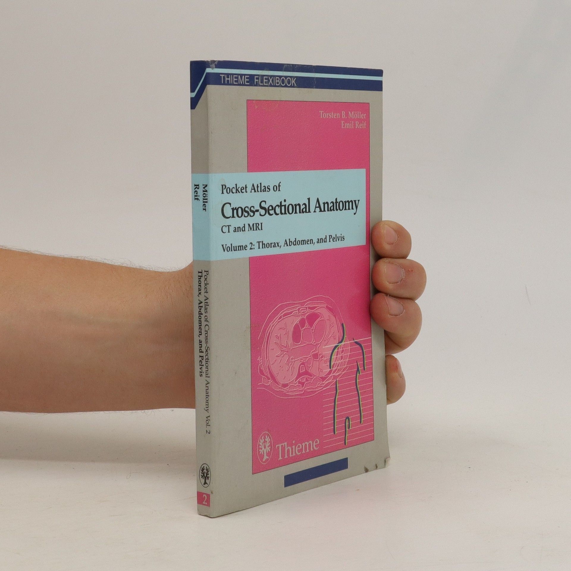
The second of a two volume set which describes the anatomical details visualized in diagnostic tomography. As a comprehensive reference it is an aid when interpreting images for anatomic structures presented in representative cross-sectional CT and MRI images.
Atlas of sectional anatomy
The Musculoskeletal System
A handy, full-color resource for interpreting musculoskeletal MRI scans with confidenceThis superbly illustrated atlas provides a comprehensive presentation of the normal sectional anatomy of the musculoskeletal system to aid in the diagnosis of diseases affecting the joints, soft tissues, bones, and bone marrow. A precise, full-color drawing accompanies each high-quality sectional image, helping the reader to gain a solid understanding of the topographic anatomy and to differentiate between normal and pathologic conditions. Following examples of whole-body imaging, the atlas offers complete representations of the spinal column and the upper and lower extremities. The contiguous images of the extremities in transverse sections facilitate the identification of structures extending beyond the joints. Key features: Top-quality MRI scans, including whole-body views, produced with the most current, high-performance equipment Full-color illustrations drawn by the authors for optimal precision and accuracy Easy identification of anatomic structures through a uniform color code in the drawings Contiguous cross-sectional anatomy of the extremities Information on the location and direction of each slice for rapid orientation Atlas of Sectional Anatomy: The Musculoskeletal System is an invaluable reference for the daily practice of radiologists, radiology residents, and radiologic technologists.

Pocket atlas of radiographic positioning
- 378 pages
- 14 hours of reading
A practical how-to guide that provides detailed information on obtaining high-quality radiographic images for optimal evaluation and interpretation of normal, abnormal, and pathological anatomic findings. It shows positioning techniques for all standard examinations in conventional radiology, CT and MRI.
Normal findings in CT and MRI
- 250 pages
- 9 hours of reading
The key for any beginning radiologist who wishes to recognize pathological findings is to first acquire an ability to distinguish them from normal ones. This outstanding guide gives beginning radiologists the tools they need to systematically approach and recognize normal MR and CT images. Highlights include: Reference-quality images from the author's own teaching files show all standard normal findings as seen in CT and MRI Checklists in each section offer the reader a systematic way to approach the images Thorough guidelines to help beginning radiologists dictate their reports Lists of standard measurements and tips for ruling out pathology
MRI – Magnetic Resonance Imaging – is now established as a valuable tool in diagnosis, treatment and monitoring of disorders of bone, joint and muscle. MRI will become the diagnostic procedure of choice: unlike conventional radiology, it is harmless, and the new fast MRI machines are small, light, cheap and easy to use. This comprehensive volume demonstrates the normal and abnormal anatomy with MRI images complemented by corresponding line diagrams in full colour, drawn by the authors from the scans for greatest accuracy.
MRI parameters and positioning
- 341 pages
- 12 hours of reading
Packed with information on the practical aspects of MRI, this user-friendly text covers everything from advice on optimal positioning of patients to recommendations for setting the appropriate scanning parameters. Each consistently organized chapter follows the chronology of a standard procedure - the authors present essential information on preparation and necessary materials first. Then they skilfully guide the readers through special considerations in positioning and coil selection, protocols for conducting the exam, examples of various sequences, and possible modifications. Numerous tips, tricks, and pointers explain how to avoid potential complications. Highlights of the second edition: 340 high-quality MRI scans and anatomical drawings New and expanded sections on MR angiography of pulmonary arteries and pelvic and leg vessels; the CARE Bolus Technique; whole-body MRI; and more Information on the latest protocols for MR urography, cholangiography, and colonography Consistent chapter structure for maximum accessibility on the job and at the MRI workstation Each section contains plenty of space on each page for personal notes A guide to the most important MRI studies, the second edition of MRI Parameters and Positioning is an indispensable companion for all radiologists, radiology residents, and radiologic technologists.